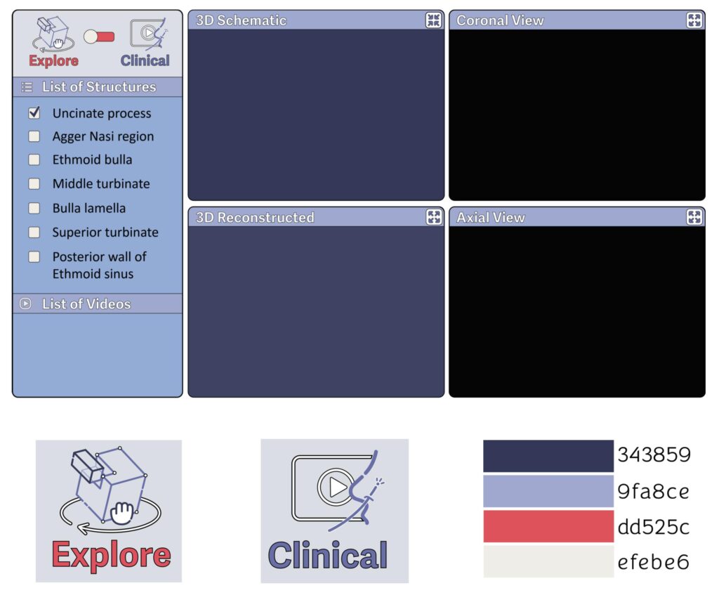
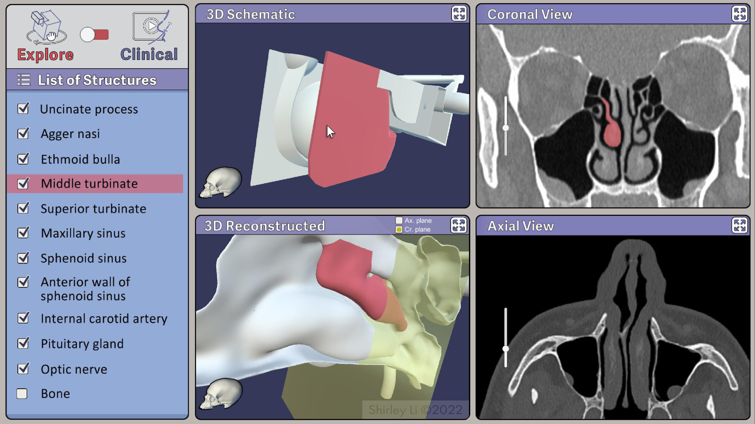
Master’s Thesis

icon thesis

Exploring the Sinonasal Cavity in Three Dimensions:

Teaching Otolaryngology Surgical Trainees Clinical Anatomy Using a Web-based Learning Resource

Understanding the three-dimensional (3D) anatomy of the nasal cavity, paranasal sinuses, skull base, and their surrounding structures is essential to perform safe and efficient endoscopic sinus surgery.

However, the learning curve to understand these complex anatomic relationships is steep. Unfamiliarity with 3D spatial relationships between structures and the failure to identify relevant anatomical landmarks can lead to intraoperative disorientation, which poses risk to patients.

Currently, there does not exist an effective teaching tool at the junior resident or medical student level.

Most of the teaching resources are limited to 2D visualization that poorly communicates the spatial relationships between structures in this anatomic region.

Although 3D visualization has been explored to develop clinical training simulations and CT-derived 3D models been used for clinical education, it has never been used to create teaching tool that allows learners to see and manipulate sinonasal structures in 3D. 


To address the need for improved teaching resource for otolaryngology surgical trainees, the objective of this thesis is to create a web-based interactive resource that aimed to use a 3D schematic model to teach clinical sinonasal anatomy and subsequently improve surgical training outcome. 

This resource will offer a comprehensive and multidimensional visualization of the sinonasal cavity that will allow learners to explore structures in 3D and help bridge gaps between different types of clinical visualization.

In this project, a 3D schematic model was created using Cinema 4D to highlight the spatial relationship of important anatomic landmarks.

A patient CT dataset was obtained (IRB approved) and segmented. The mesh was then polished using ZBrush to generate a CT-reconstructed 3D model.

Adobe Photoshop was used to process and label CT images, and Adobe Illustrator was used to create 2D assets for user interface. Finally, the 3D models and 2D visualizations were integrated into an interactive application using Unity.

3D Schematic Model

User Interface

Two Learning Modes

The Explore Mode

The fully manipulatable 3D schematic model is presented alongside a 3D CT-reconstructed model and 2D CT visualizations in axial and coronal plane. The two 3D models can be manipulated in concert, structures can be highlighted and turned off from the navigation menu on the left.
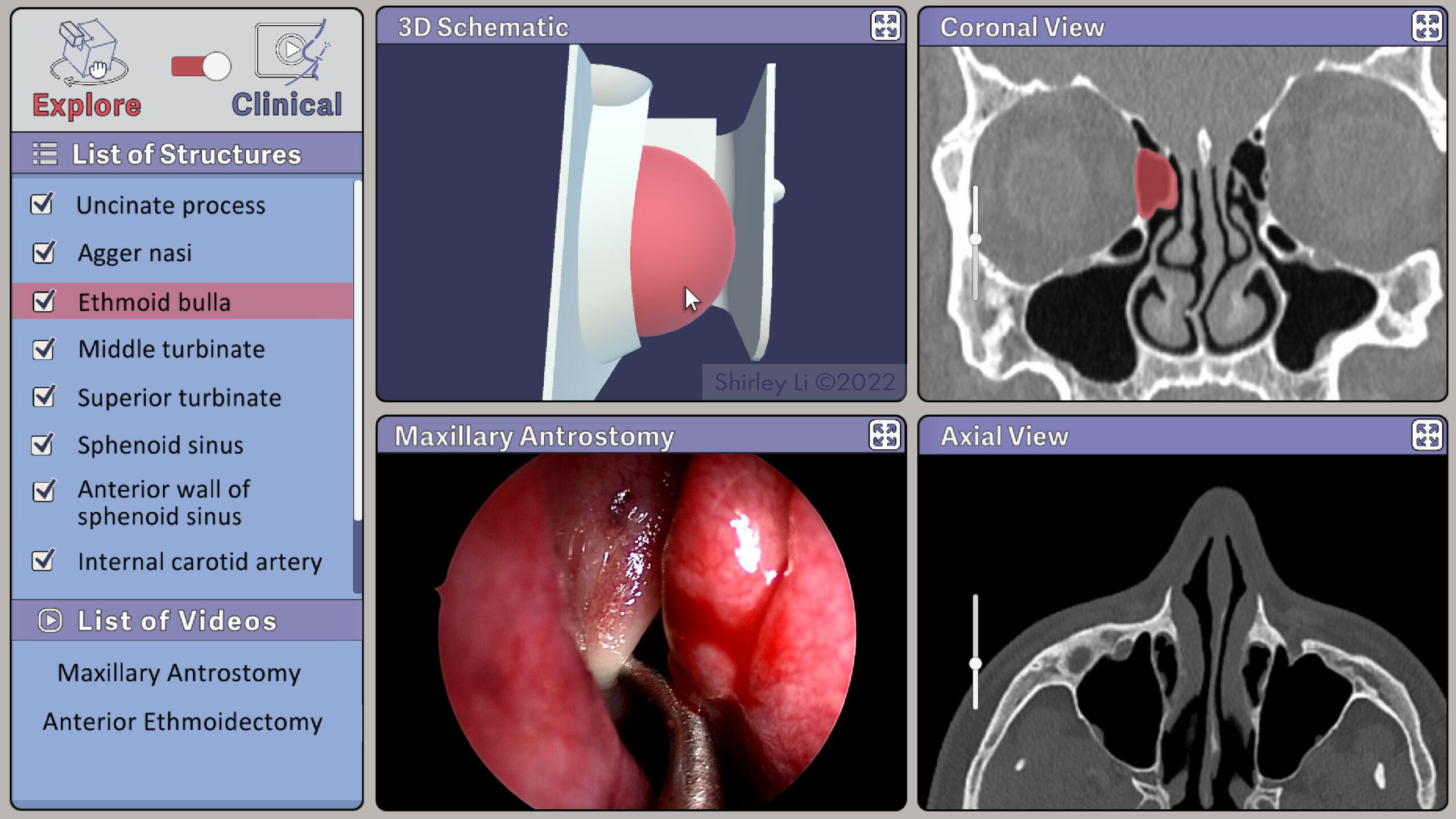
The Clinical Mode

The 3D schematic model is presented alongside a panel featuring intraoperative video. The range of rotation of the schematic model will be limited to what the intraoperative endoscopic angle of view would be to mimic surgical perspective.

Demo clip:

Achievements

Future Directions